Hoencamp et al., 2021 - 3D genomics across the tree of life reveals condensin II as a determinant of architecture type - Media Kit
-
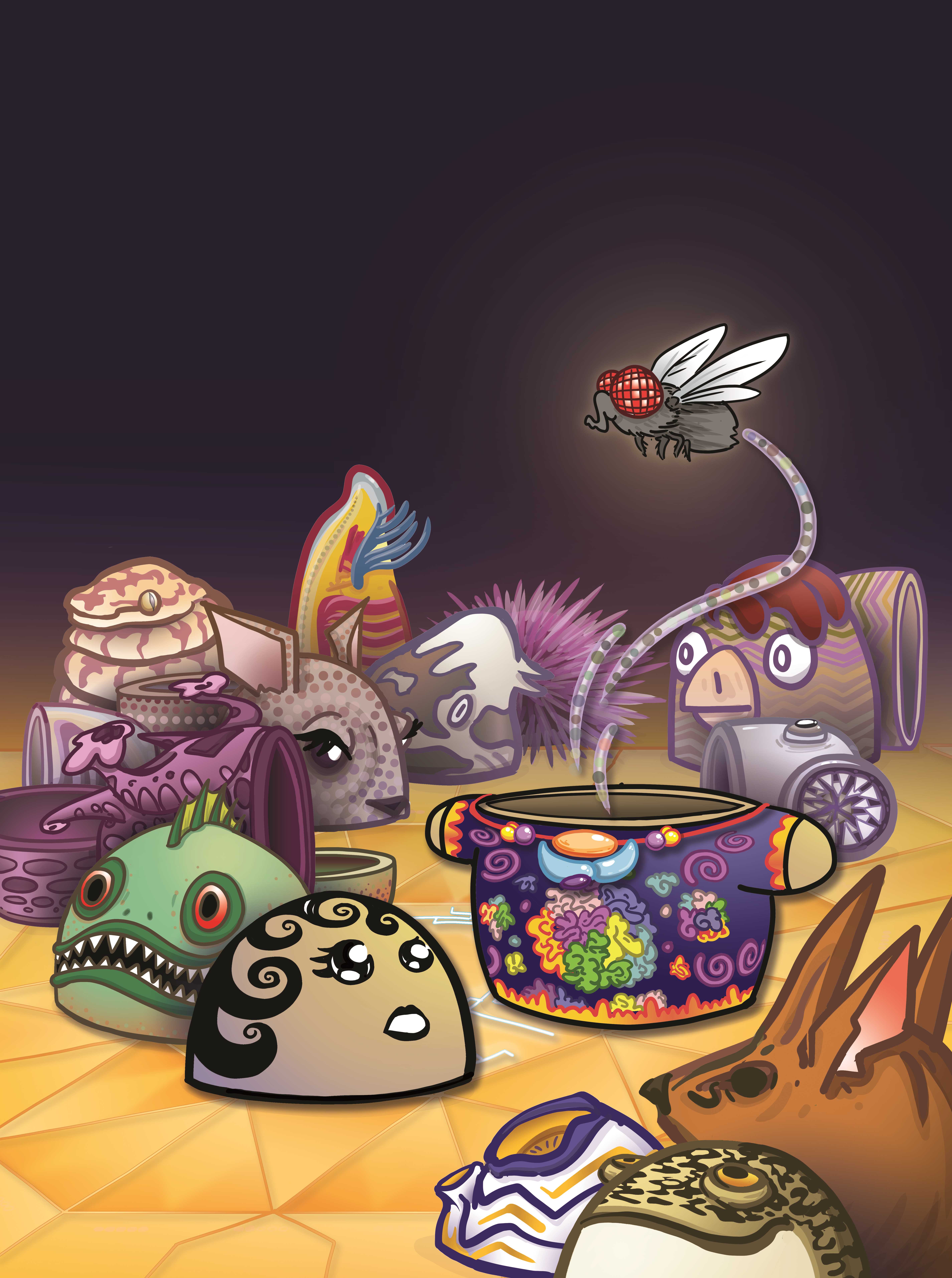
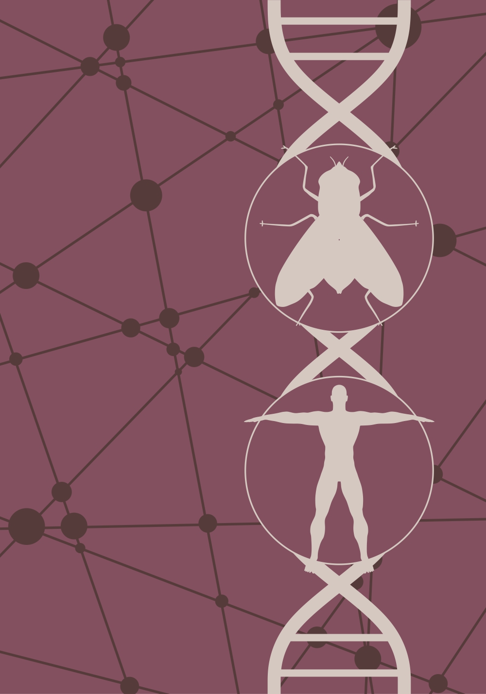
The inner fly
Artist: Adam Fotos
Image Credit: Adam FotosThis image depicts, in the form of stacking dolls, a number of species examined in our paper. We analyze and group the species by their nuclear architecture type, and transform a typical human architecture into a typical fly nuclear arrangement in a condensin II disruption experiment.
-
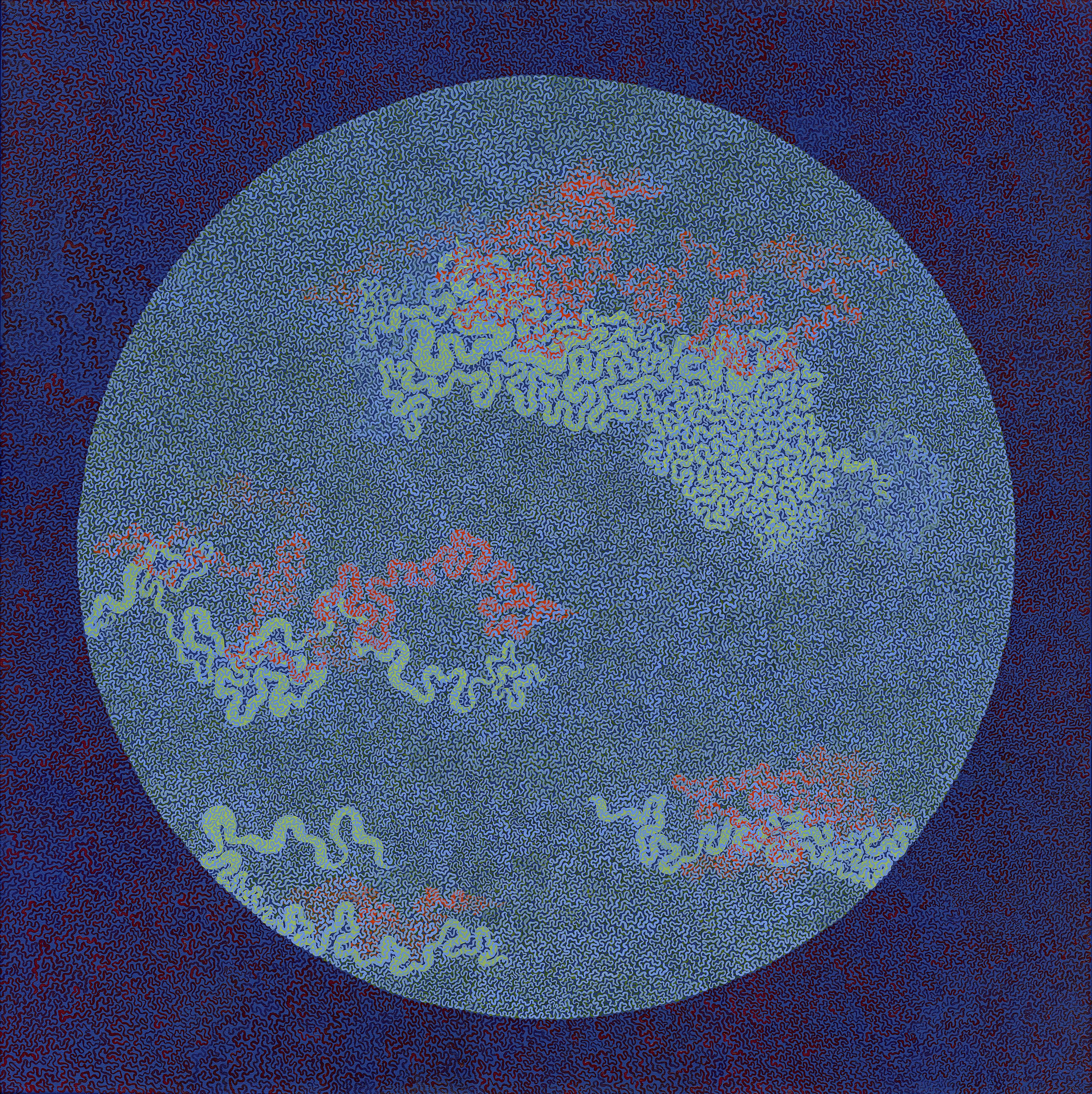
Nucleus, 2017
Artist: Mary Ellen Scherl
Image Credit: Mary Ellen Scherl
Medium: Acrylic on canvas
Dimensions: 34 x 34 in.An artist’s interpretation of chromatin folded up inside the nucleus. The artist has rendered an extraordinarly long contour into a small area, in two dimensions, by hand.
-
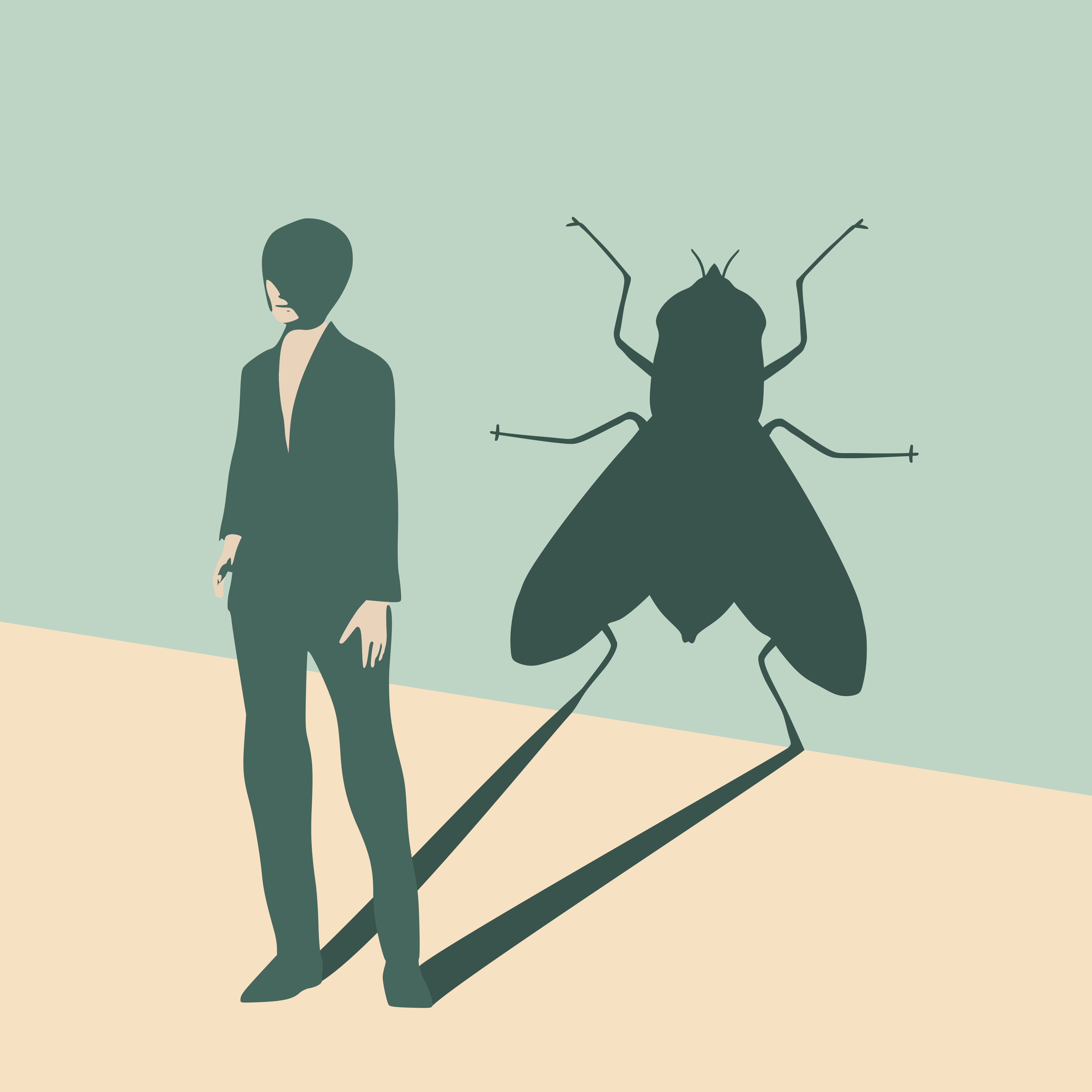
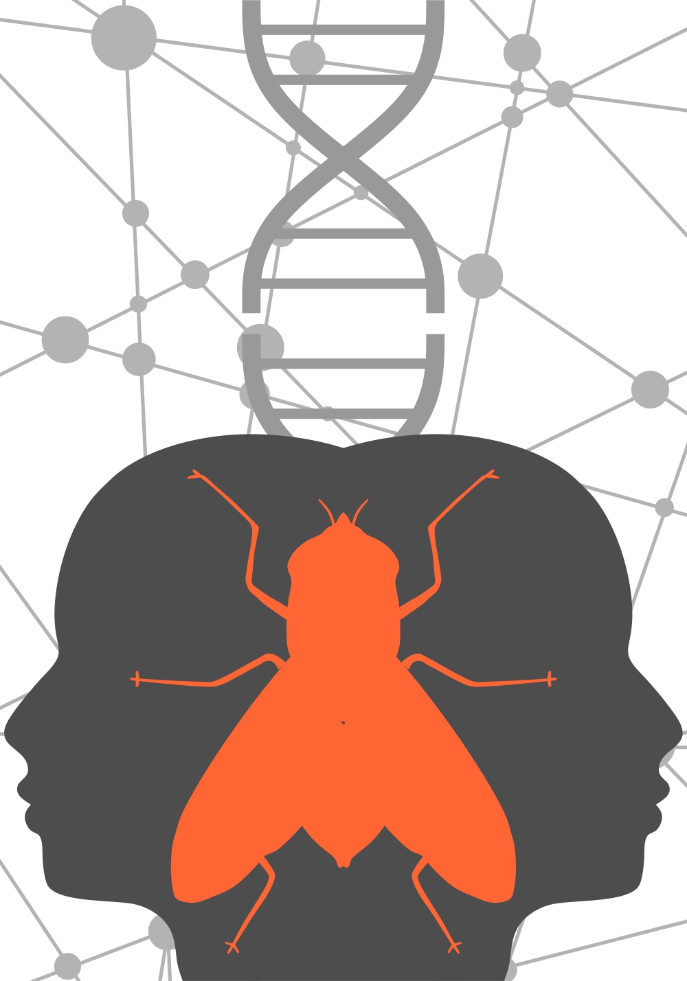
The fly shadow
Artist: Evgeny Gromov
Image Credit: Evgeny GromovThis image depicts a human casting a fly shadow. In our paper we show that we can transform the nuclear arrangement in a human cell into that typical of a fly nucleus.
-
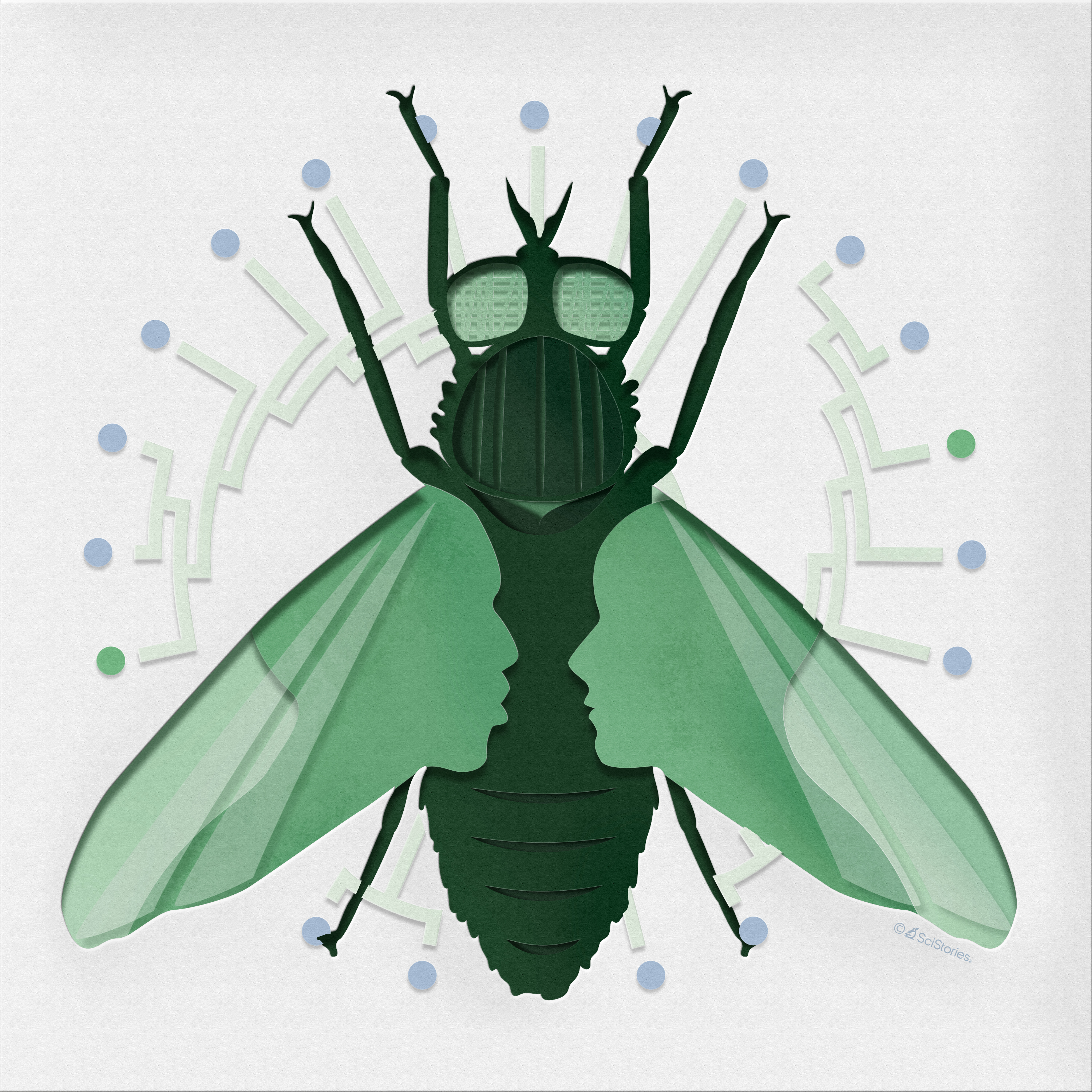
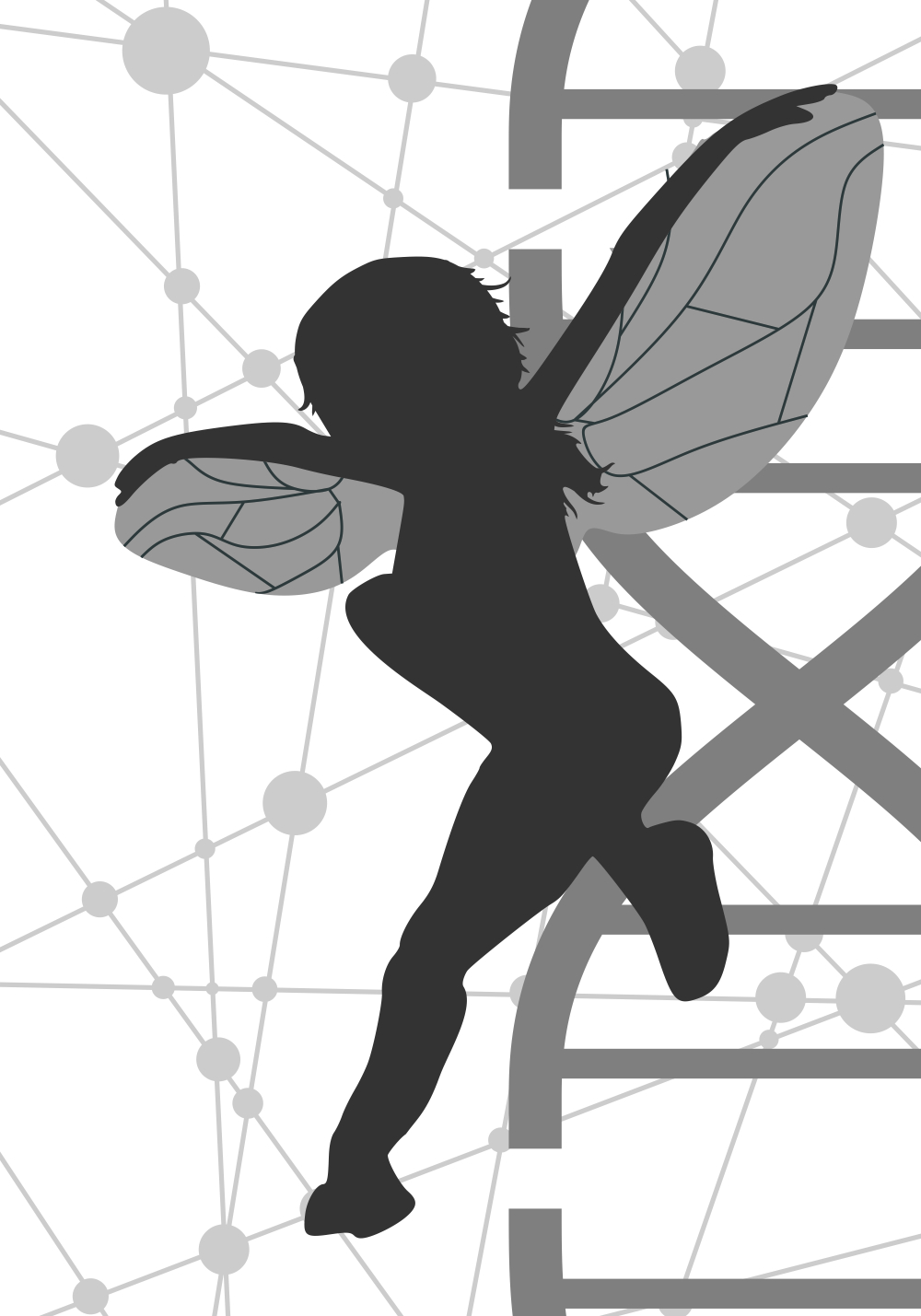
The Fly III
Artist: Artwork by SciStories LLC, https://scistories.com
This work stemmed from a discussion of an optical illusion that we, in a way, create in the Hoencamp et al. manuscript where, by disrupting condensin II protein, we create human cells that resemble those of a fruit fly.
-
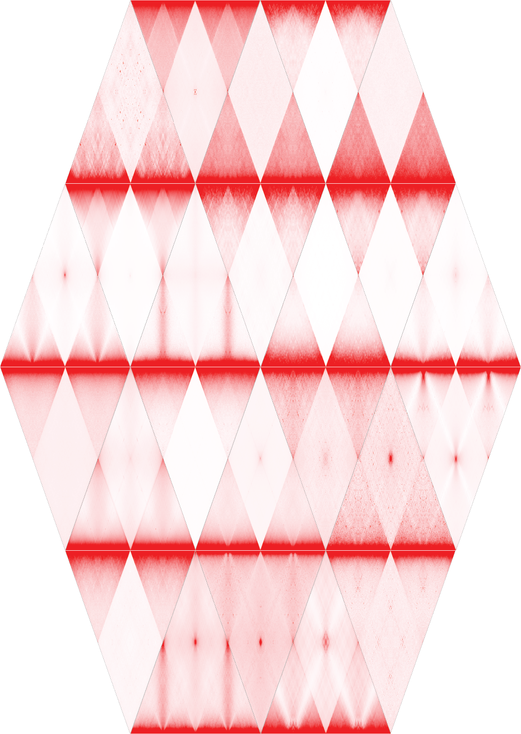
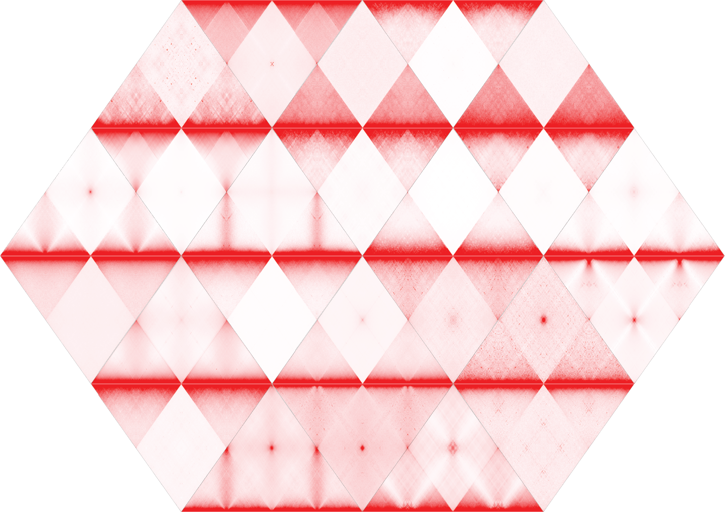
Nuclear Tilings
Image Credit: Olga Dudchenko, Erez Lieberman Aiden
This image shows an arrangement of 24 contact maps from 24 different organisms from across the tree of life. The contact maps show how chromosomes in different species interact with each other, highlighting the different contact features such as chromosome territories, centromere-to-centromere and telomere-to-telomere contacts and foldback along the centormere-to-telomere axis.
-

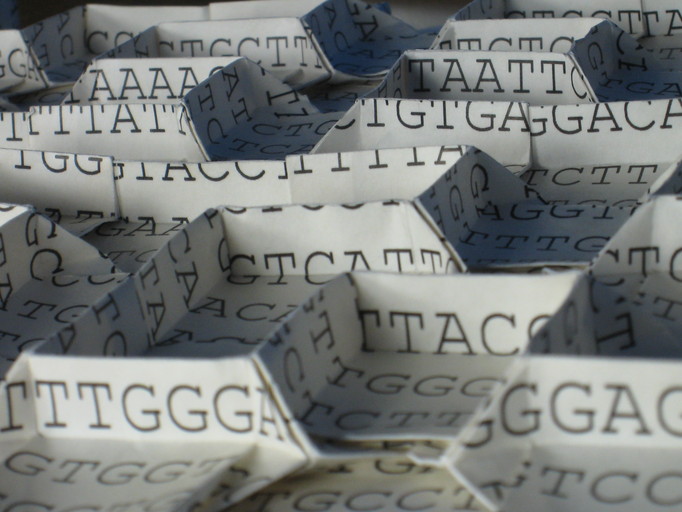
Chromosome origami
Image Credit: Jason Ku, Erik Demaine
Two photographs showing a sequence of human chromosome 14 folded into a three-dimensional pattern. In our work we study how the genomes of different organisms across the tree of life fold in 3D.
-
Chromosome origami
Image Credit: Jason Ku, Erik Demaine
Two photographs showing a sequence of human chromosome 14 folded into a three-dimensional pattern. In our work we study how the genomes of different organisms across the tree of life fold in 3D.
-
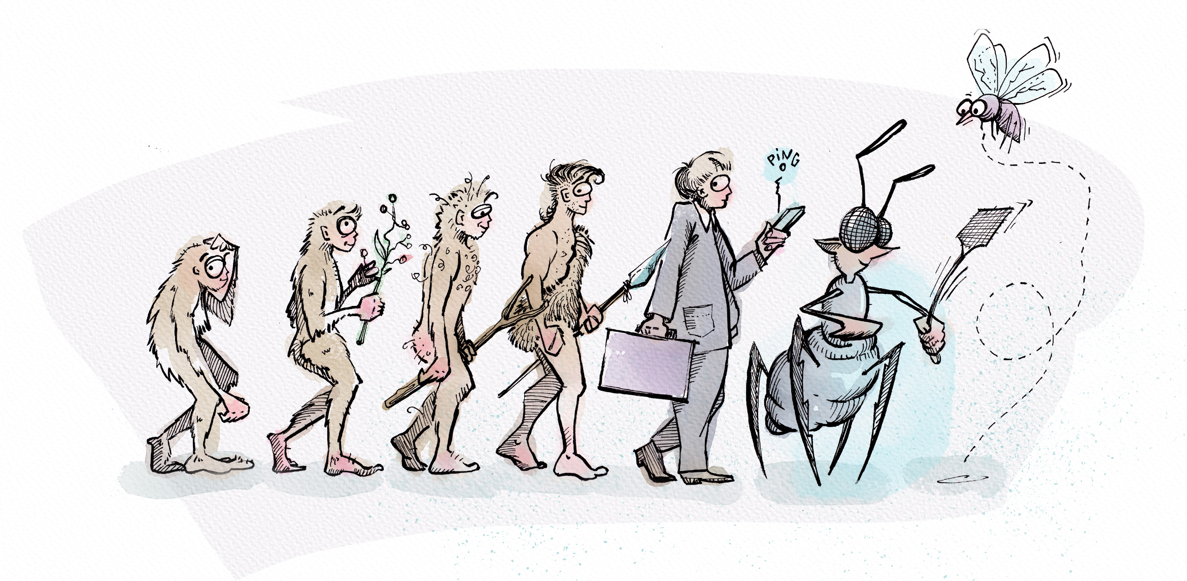
Evolution!?
Artist: Joris Koster
Image credit: ©Netherlands Cancer Institute/Joris Koster Artwork
Dimensions: 11,7 x 5,7 inch (rectangular) and 11,7x11,7 inch (square)An artist’s interpretation of evolution from primates, via modern humans to mosquitoes. This artwork is a play on our data in which we show that we can change the organization of the human genome into something that more resembles the genome organization of mosquitoes.
Evolution_rectangle.psd -
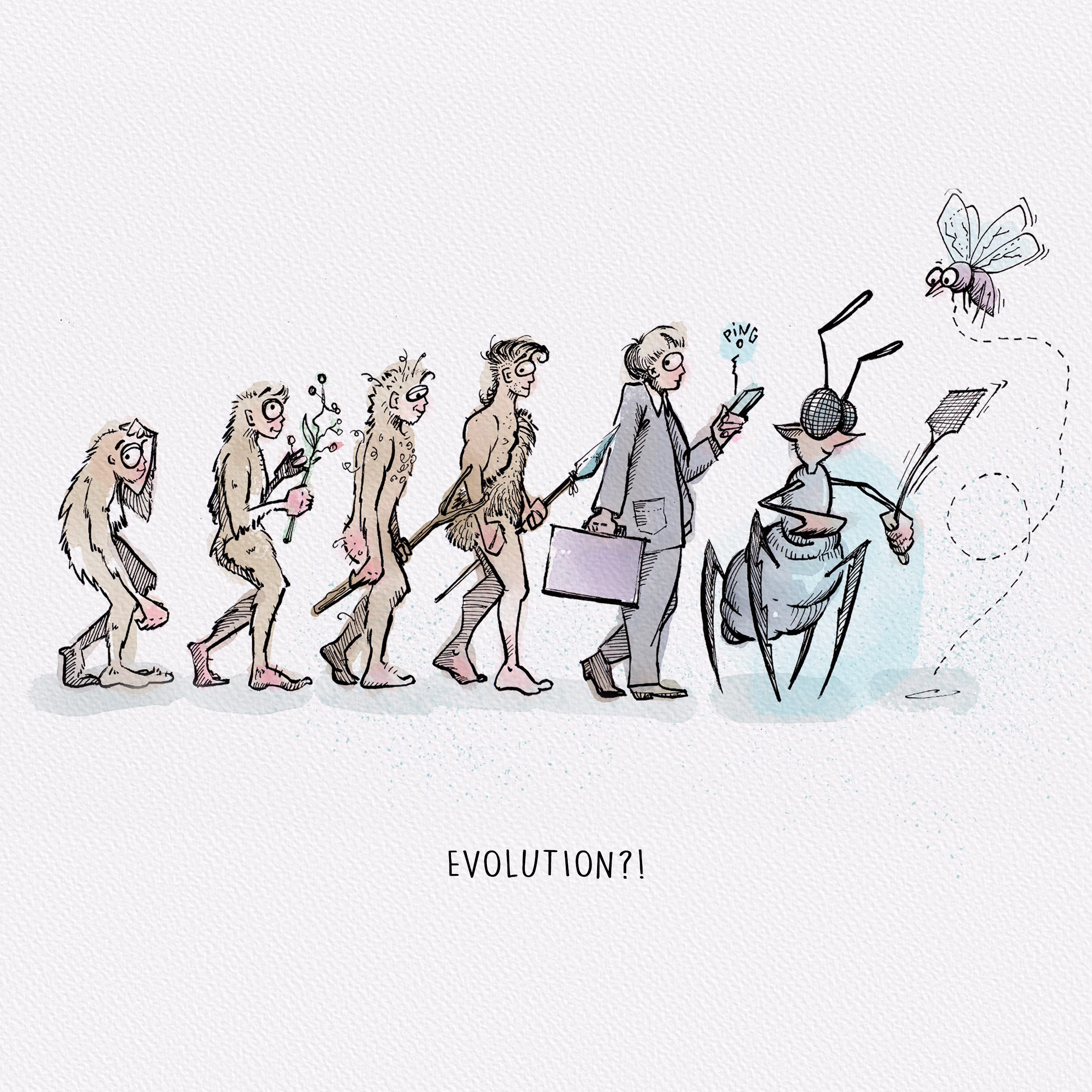
Evolution!?
Artist: Joris Koster
Image credit: ©Netherlands Cancer Institute/Joris Koster Artwork
Dimensions: 11,7 x 5,7 inch (rectangular) and 11,7x11,7 inch (square)An artist’s interpretation of evolution from primates, via modern humans to mosquitoes. This artwork is a play on our data in which we show that we can change the organization of the human genome into something that more resembles the genome organization of mosquitoes.
Evolution_square.psd -
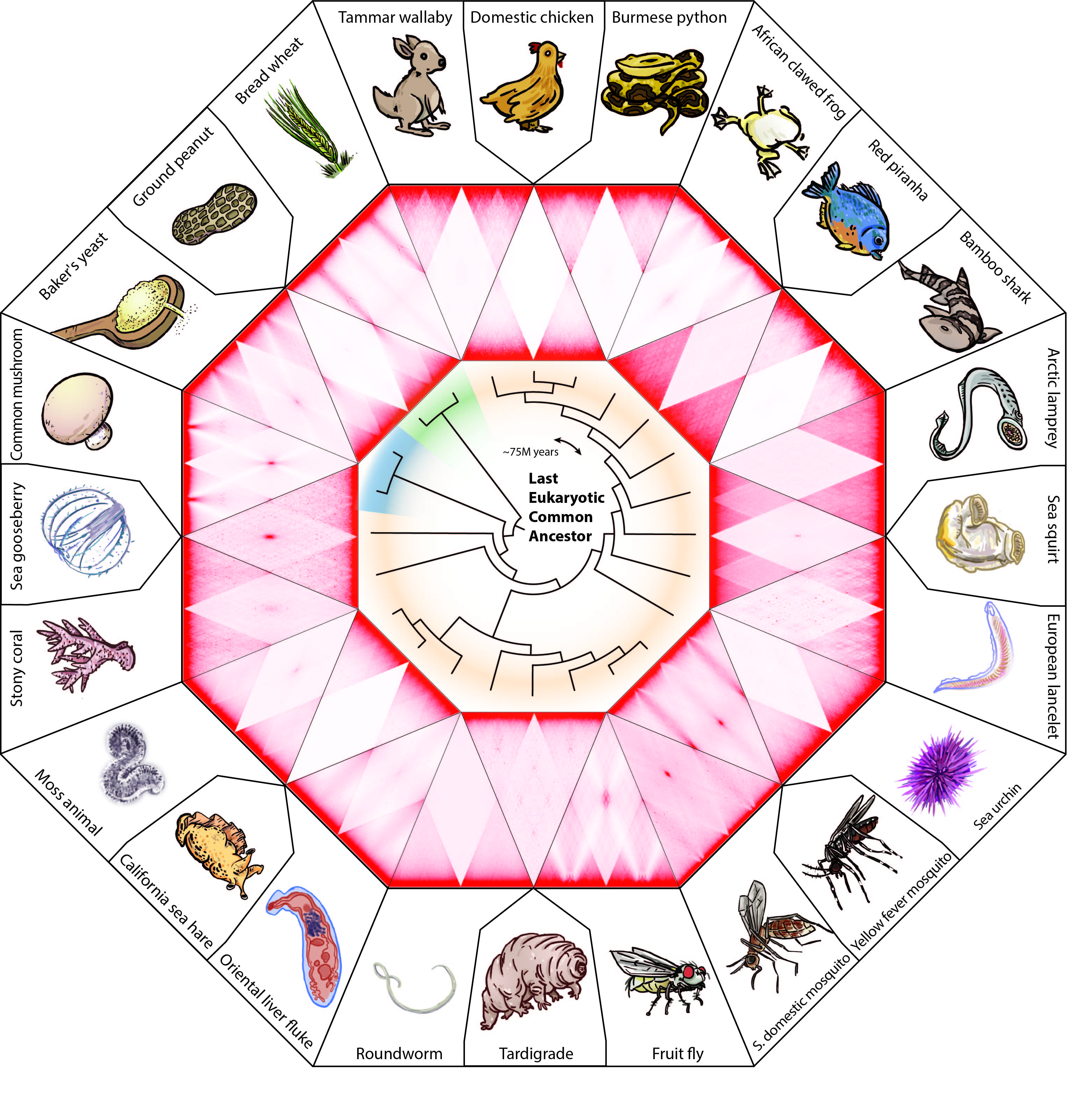
A comprehensive overview of genome organization across eukaryotic evolution
Image credit: Adam Fotos, Olga Dudchenko, Benjamin Rowland, Erez Aiden
A simplified version of Figure 1 from the Hoencamp et al., 2021 paper showing the menagerie of chromosome contact patterns in nuclei of various animals and plants.
-

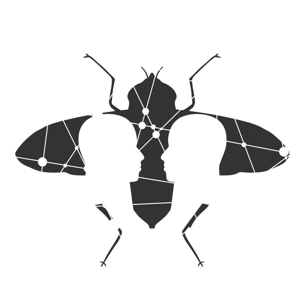
Artist's interpretation, Evgeny Gromov 1
Artist: Evgeny Gromov
Image Credit: Evgeny Gromov -

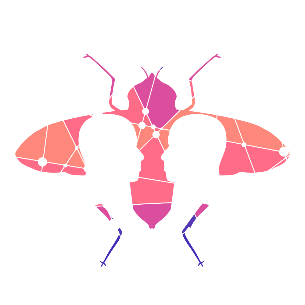
Artist's interpretation, Evgeny Gromov 2
Artist: Evgeny Gromov
Image Credit: Evgeny Gromov -
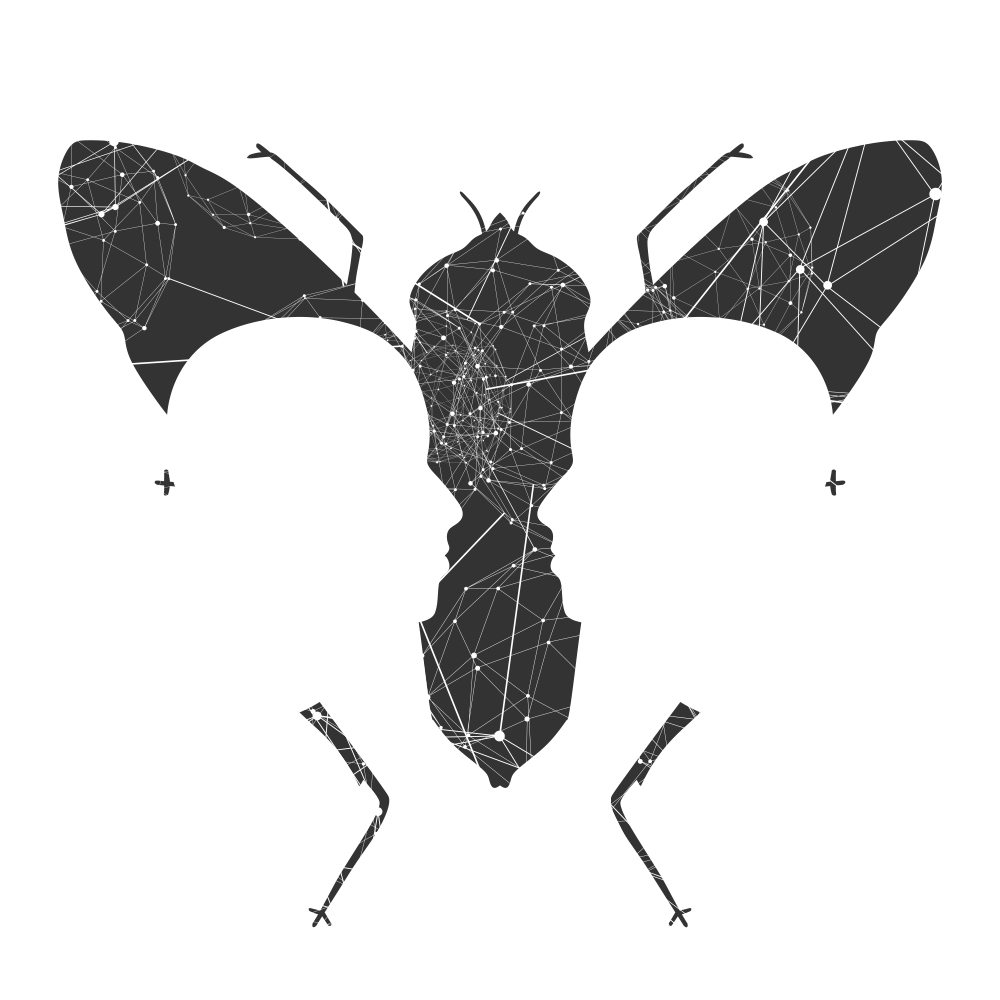
Artist's interpretation, Evgeny Gromov 3
Artist: Evgeny Gromov
Image Credit: Evgeny Gromov -
Artist's interpretation, Evgeny Gromov 4
Artist: Evgeny Gromov
Image Credit: Evgeny Gromov -
Artist's interpretation, Evgeny Gromov 5
Artist: Evgeny Gromov
Image Credit: Evgeny Gromov -

Artist's interpretation, Evgeny Gromov 6
Artist: Evgeny Gromov
Image Credit: Evgeny Gromov -
Artist's interpretation, Evgeny Gromov 7
Artist: Evgeny Gromov
Image Credit: Evgeny Gromov -
Artist's interpretation, Evgeny Gromov 8
Artist: Evgeny Gromov
Image Credit: Evgeny Gromov -
Artist's interpretation, Evgeny Gromov 9
Artist: Evgeny Gromov
Image Credit: Evgeny Gromov -
Artist's interpretation, Evgeny Gromov 10
Artist: Evgeny Gromov
Image Credit: Evgeny Gromov -
Artist's interpretation, Evgeny Gromov 11
Artist: Evgeny Gromov
Image Credit: Evgeny Gromov -
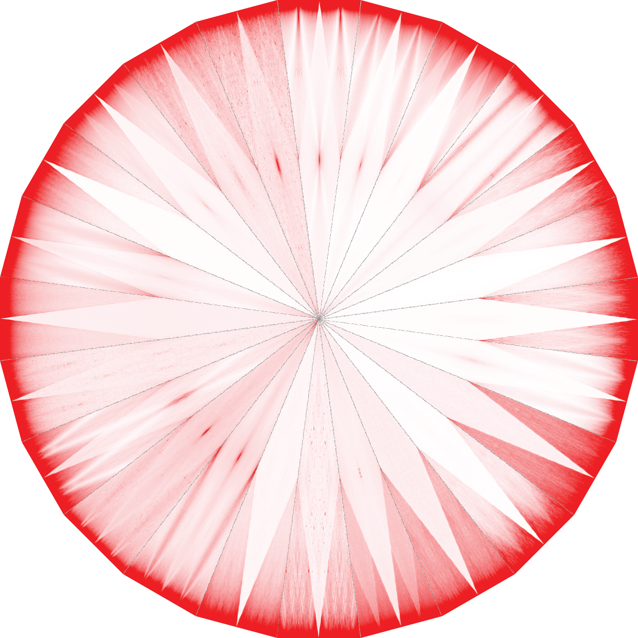
Nuclear Tilings 2
Image Credit: Olga Dudchenko, Erez Lieberman Aiden
This image shows an arrangement of 24 contact maps from 24 different organisms from across the tree of life. The contact maps show how chromosomes in different species interact with each other, highlighting the different contact features such as chromosome territories, centromere-to-centromere and telomere-to-telomere contacts and foldback along the centormere-to-telomere axis.
-
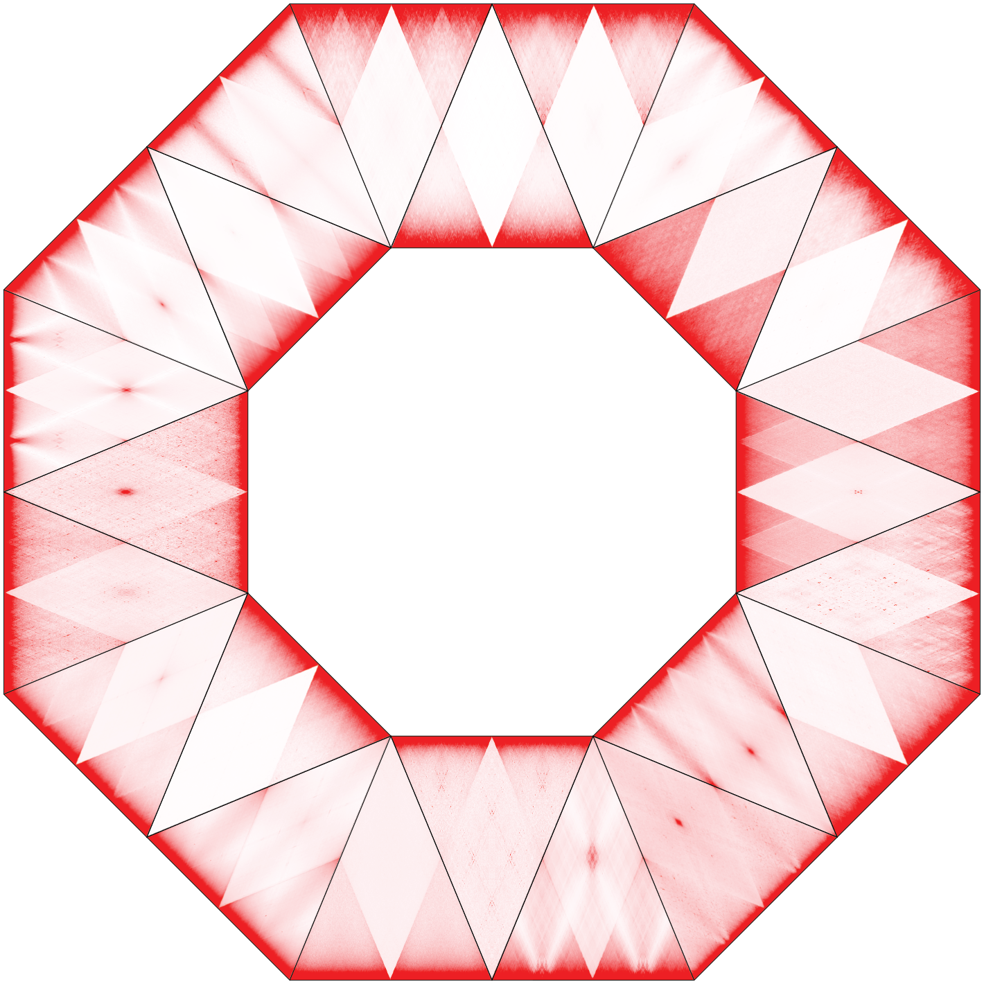
Nuclear Tilings 3
Image Credit: Olga Dudchenko, Erez Lieberman Aiden
This image shows an arrangement of 24 contact maps from 24 different organisms from across the tree of life. The contact maps show how chromosomes in different species interact with each other, highlighting the different contact features such as chromosome territories, centromere-to-centromere and telomere-to-telomere contacts and foldback along the centormere-to-telomere axis.
-
Nuclear Tilings 4
Image Credit: Olga Dudchenko, Erez Lieberman Aiden
This image shows an arrangement of 24 contact maps from 24 different organisms from across the tree of life. The contact maps show how chromosomes in different species interact with each other, highlighting the different contact features such as chromosome territories, centromere-to-centromere and telomere-to-telomere contacts and foldback along the centormere-to-telomere axis.